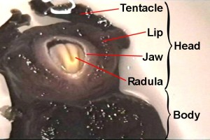
Supplementary Material

Operant reward learning in Aplysia: Neuronal correlates and mechanisms
Björn Brembs, Fred D. Lorenzetti, Fredy D. Reyes, Douglas A. Baxter, John H. Byrne
(HTML, PDF)

Supplemental Video. Aplysia biting behavior. The consummatory phase of Aplysia feeding behavior (biting) occurs in an all-or-nothing fashion and is unambiguously quantifiable (1, 2). It consists of four phases: jaw opening, odontophore/radula protraction in the open state, odontophore/radula retraction in the closed state and jaw closure. Biting occurs spontaneously as well as reflexively, in nature as well as in the laboratory. If food is present, it leads to the ingestion of food through the buccal cavity. In this 8 s video sequence, the animal has positioned itself under the water surface (as they often do). Its tentacles (anterior) are at the top of the screen. The sequence contains the opening of the jaws, followed by the protraction of the radula (cream colored tongue-like organ) in the open state, the closure of the radula, the retraction of the radula in the closed state and the closing of the jaws.

To view this video, download a QuickTime viewer.

Quicktime video: 176x144 pixels (123,925 bytes) | 800x600 pixels (2,602,794 bytes)


Supplemental Methods 1. Surgical procedures and in vivo recordings. Aplysia californica (100-200 g) were obtained from Alacrity Marine Biological Specimens (Redondo Beach, CA) and Marinus (Long Beach, CA). They were kept individually in rectangular perforated plastic cages floating in aerated artificial seawater (Instant Ocean; Aquarium Systems, Mentor, OH) at a temperature of 12-15°C. Animals were fed ~1 g of dried laver, 3 times a week. To help ensure that all animals were in a similar motivational state, experimental animals were food deprived 3-5 days before surgery.
Extracellular electrodes were prepared from 25.4 µm insulated stainless steel wire (304 TRI-ML, California Fine Wire Co., Grover Beach, CA) by scraping the insulation from 2 mm at one end of a piece (~30 cm) of wire and from about 15 mm at the other end. Two such wires were used, one for recording/stimulating, the other for reference. The two long de-insulated ends were soldered to gold contacts, whereas one of the short de-insulated ends was fashioned into a small hook for placement on the nerve (for stimulation/recording). The other wire served as the reference electrode and its tip was left straight.
Just prior to surgery, the animals were fed a single bite of seaweed, to probe the motivational state, overall feeding behavior and health of the animals. Animals that did not feed were discarded. Each animal was then anaesthetized by injecting isotonic MgCl2 (360 mM) solution (30% body weight) into the hemolymph and transferred onto a block of ice (made of seawater), covered with artificial seawater. The animal was positioned with its left body side up and two hooks shaped from hypodermic needles (30 G1/2; Becton Dickinson, Franklin Lakes, NJ) were placed in the skin ~3 cm apart ~1.5 cm ventral of the eye and in parallel with the anterior-posterior axis of the animal. The hooks were fastened by threads on either end of the tank and tightened so as to lift the fold of skin between them above the water surface. A small (1-1.5 cm) incision was made between the hooks alongside the fold and with the eye being at the height of the anterior third of the incision. During the surgery, the thread suspended hooks were used to keep the wound above the water level to prevent leakage of hemolymph, or seawater from entering the animal. The incision was kept open using a second pair of hooks arranged perpendicularly to the first pair. A moveable support was used to lift the buccal mass and expose the buccal ganglia and their nerves. Additional manipulators were then used to place the hook-electrode around the anterior branch of the esophageal nerve (En2), close to the anterior/posterior branch point. The electrode was secured and insulated by a drop of superglue (Loctite Quick Gel, part # SGG-2B, Loctite, Rocky Hill, CT). After inserting the reference electrode to float free in the hemocoel, the animal was closed using 4-6 stitches of 4-0 black braided silk suture (Ethicon, Somerville, NJ), with the electrodes exiting the animal at the posterior end of the incision. The entire procedure lasted between 45-60 minutes for each animal. After the surgery, the animals were placed into individual rectangular perforated plastic cages and left to recover overnight at 12-15° C. Electrode signals were amplified using a differential AC amplifier (model 1700; A-M Systems, Everett, WA), filtered by a 100 Hz low cut-off filter and a 1 kHz high cut-off filter. One day after surgery, extracellular activity in the anterior branch of the esophageal nerve was recorded while the animal was observed in a round glass bowl (radius: 80 mm, depth: 70 mm) placed on a mirror. During the observation period, the animal was stimulated to bite and swallow with pieces of seaweed. The animals were not restrained and the length of the wires permitted a full range of motion in the bowl. Animals used for in vivo recording were not used in the conditioning studies.


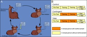
Supplemental Methods 2. Operant reward learning. The occurrence of spontaneous bites is dependent on a number of variables. While it can be observed in freshly caught specimens, it is comparatively rare. To successfully conduct experiments, certain measures need to be taken to increase the frequency at which this behavior occurs. To help ensure that all animals were in a similar motivational state, experimental animals were food deprived 3-5 days before surgery. One day after implanting the stimulating electrodes, the animals were fed a single bite of seaweed 30 minutes before the experiment to motivate the animal to search for food. Seaweed extract was prepared by incubating a 10 cm x 20 cm piece of seaweed in 300 ml of artificial seawater for 30 minutes. Pilot studies found seaweed extract to increase the overall probability of biting behavior to occur. Just prior to the experiment, 50 ml of the supernatant were added to 400 ml of fresh artificial seawater. The animal was then transferred into a round glass bowl (radius: 80 mm, depth: 70 mm) containing these 450 ml of diluted seaweed extract and the bowl placed on a mirror to be able to better observe the animal. The experiment was performed in a climate chamber at 15° C and 60% rel. humidity. Unrestrained in the bowl, the animal moved around freely and engaged in spontaneous behaviors (A). Throughout the experiment, the animal was observed and all bites recorded. A bite (see supplemental video) was defined as opening of the jaws and protraction of the radula. Before the start of the experiment, animals were assigned to one of three groups (B): i) a control group that did not receive any stimulation, ii) a contingent reinforcement group which received En2 stimulation whenever the jaws closed after a bite during training, or iii) a yoked control group that received the same sequence of stimulations as the contingent group, but the stimulation occurred uncorrelated with their behavior. Except for the different reinforcement schedules, all animals in all three groups were treated identically. Application of reinforcement was the only difference between training and test. In the early phase of the study, the animals were assigned randomly to each group. As the study progressed, the animals were assigned to each group so as to balance pre-test bite rate between groups. Experimental sessions consisted of four consecutive five-minute periods. In each period, the number of bites was recorded. The final test period was either immediately after training (I-Test) or 24 h after the beginning of the experiment (L-Test). A Grass S48D stimulator (Grass Instruments, Quincy, MA) generated 10 ms pulses for extracellular nerve stimulation (30 Hz, 3 s). Pilot studies determined that a suitable intensity of the stimulation was 8 V. At this voltage, usually no behavioral response could be observed. Occasionally, an animal (mostly yoked controls) would show a jaw opening without radula protraction or a rejection-like behavior (i.e., the radula appeared to be closed during protraction) to the first few stimulations only. Such responses were never observed spontaneously. If they met the definition of a bite (i.e., the radula was protracted), they were scored as bites irrespective of the subjective impression of the observer. Animals that were tested after 24 h spent the time between training and test in individual rectangular perforated plastic cages in aerated artificial seawater at 12-15° C. On the next day, the animals were placed back into the glass bowl with seaweed extract, but without being fed before the test. After the experiments, all animals were sacrificed and electrode placement verified. Only animals that produced 0 < n < 31 bites in the pre-test were used. Several experimenters independently replicated this experiment and all their data were pooled.


Medium version | Full size version


Supplemental Methods 3. Biophysical correlates of the operant memory in B51. Animals from a second behavioral study were anesthetized by injecting a volume of isotonic MgCl2 equivalent to 50% of the animal's weight. Buccal ganglia were removed and pinned on a Sylgard-coated Petri dish containing artificial seawater (ASW). The composition of the ASW was: 450 mM NaCl, 10 mM KCl, 30 mM MgCl2(6H2O), 20 mM MgSO4, 10 mM CaCl2(2H2O), 10 mM HEPES, with pH adjusted to 7.4. The ganglion ipsilateral to the esophageal nerve stimulation was desheathed on the rostral side. Desheathing was performed in the presence of high divalent cation ASW solution, which contained concentrations of CaCl2 and MgCl2 that were three times the normal level. Osmolarity was maintained by correspondingly decreasing the concentration of NaCl. After desheathing, the medium was changed to normal ASW.
Neuron B51 was identified based on its relative size and position in the ganglia. The identity of the cell was confirmed by its pattern of electrical activity during a buccal motor program, membrane properties, and its characteristic plateau potential as described in (3,4). Recording temperature was 15° C.
Conventional two-electrode current-clamp techniques were used for intracellular recordings (Axoclamp-2A, Axon Instruments, Burlingame, CA). Fine-tipped glass microelectrodes (resistance 10-15 MOhms) were filled with 2 M potassium acetate. The cell was current-clamped at -60 mV for the duration of the experiment. Five minutes after impalement with the electrodes, the input resistance and the burst threshold were measured. If a spontaneous motor pattern occurred during testing, the test was halted and then resumed 1 minute after the cessation of the pattern. The input resistance of B51 was determined by injecting a hyperpolarizing current of 5 nA for 5 s. The burst threshold of B51 was defined as the minimum amount of depolarizing current necessary to elicit activity in B51 that outlasted the current pulse. The burst threshold was tested by a series of successively higher amplitude depolarizing current pulses (in 1 nA increments) with a duration of 5 s. The series was spaced with 10 s between the end of one pulse and the start of another. In this way the minimum amount of current necessary to elicit a plateau potential could be determined. After these two properties were measured, the cell was released from current clamp and the resting membrane potential was determined. On average, membrane properties of B51 were recorded 100 minutes after the last training period. The experimenter performing the intracellular measurements was unaware of the experimental history of the animals.


Supplemental Methods 4. B51 cell culture and electrophysiology. Culturing procedures followed those described in (5-8). Buccal ganglia from adult Aplysia were incubated in 1% protease type IX (Sigma, St. Louis, MO) at room temperature for 24 hours and then desheathed. In pilot studies, B51 neurons in the buccal ganglia were first identified by the electrophysiological methods described previously and then dye-labelled with Fast Green (Sigma). B51 neurons were removed from the ganglia by microelectrodes with fine tips and plated on poly-L-lysine coated glass slides in petri dishes with culture medium containing 50% hemolymph and 50% isotonic L15 (Sigma). The cells were allowed to grow for 4-5 days and the medium was changed on the third day. Culture medium was exchanged for ASW prior to recording. It was found that neurite morphology coupled with the size and the relative position of the cell in the ganglia was sufficient to identify B51. Thus, these criteria were adopted as the means of identification for all the neurons used in this report.
The electrophysiological methods used to record from cultured neurons were largely the same as those used to record from neurons in the ganglia. Due to the high input resistance in cultured cells, the cells were current clamped to -80 mV. Five minutes after impalement, input resistance and burst threshold were determined. Input resistance was tested by injecting a hyperpolarizing current pulse of 0.5 nA for 5 s and burst threshold was tested in 0.1 nA increments. The cells were then divided into a contingent reinforcement and an unpaired group. Plateau potentials were generated by a 5 s long depolarizing current pulse with an amplitude 0.1 nA higher than the previously determined threshold. Both groups received 7 evenly spaced supra-threshold depolarizing current pulses in a ten-minute training period. The cells in the contingent reinforcement group received a 6 s iontophoretic pulse of dopamine immediately after the cessation of the plateau potential, whereas iontophoresis was delayed by 40 s in the unpaired group. Dopamine was iontophoresed through a fine-tipped glass microelectrode (resistance 10-15 MOhms). A retaining current of -1 nA was used during the course of the experiment. A square wave current pulse of 35 nA for 6 s was used to eject the dopamine. The concentration of dopamine in the electrode was 200 mM. An equimolar concentration of ascorbic acid was added to the electrode to reduce the oxidation of dopamine. After training, the membrane properties were measured again and compared to the pre-test levels.
Recordings were performed at room temperature.


Supplemental Discussion. From postural adaptation to social interaction, operant conditioning is one of the essential processes leading to the generation and modulation of behavior. However, its analysis has been complicated because most learning situations inseparably comprise operant and classical components. Specifically, behaving organisms constantly receive a stream of sensory input that is both dependent and independent of their behavior. The classic debate as to whether one or two processes account for the operant/classical dichotomy reflects this entanglement (e.g., 9-14). Interrupting the operant feedback loop by restraining an animal can at least partly isolate classical conditioning from the operant components. Once isolated from spontaneous behavior, in a number of systems the stimuli have been traced into the nervous system to find the point of convergence where the classical association is formed. Until now, the convergence of reinforcement and the operant behavior has remained elusive, however.
Similar to the successful isolation of the stimuli from spontaneous behavior in classical conditioning, the development of the present procedure is a critical step towards operant conditioning without any external stimuli other than the reward being contingent upon the behavior (i.e., ‘pure’ operant conditioning, 14).  So far, it can not be ruled out that the animal can perceive the sound and vibrations associated with recording the sequence of stimulations and activating the stimulator. However, given the nature of the sensory organs in Aplysia, this appears highly unlikely. Thus, in practical terms, the bite occurs spontaneously and except for the reinforcement, all external stimuli are either constant or independent of the biting behavior. The nature of the reinforcement also rules out classical contamination by predictive ‘internal’ stimuli generated by other types of reinforcement. Whereas other reinforcers like food or water need consummatory behavior (preceding the reward) to be effective, stimulation of En2 is not preceded by any other behavior or stimuli (external or internal) other than the rewarded operant behavior. It cannot be ruled out that internal stimuli are generated by the innervation of  the buccal musculature and the salivary gland by En2. However, such stimuli would be sensed after the onset of the reward and can therefore not acquire any predictive properties other than that they are not followed by reinforcement.
Contextual cues are always present during the experiment. Indeed, without contextual cues, the association would most likely have extinguished in the home tank before the 24 h test. However, these cues were constant throughout the experiment and thus are not predictive as to when exactly the reinforcement will occur (such as a classical conditioned stimulus would). Therefore, it is safe to contend that any contextual cues act as ‘occasion setters’ to indicate whenever the behavior – reinforcer relation is true and do not directly enter into the primary operant association.
With the development of in vivo and in vitro classical and operant procedures in Aplysia, the first detailed mechanistic comparison between operant and classical conditioning in the same preparation is within reach.  Ultimately, the tools now available in Aplysia will allow for studies of operant-classical interactions (e.g., 14).

References and Notes:

  1. I. Kupfermann, Behav. Biol. 10, 1 (1974).
  2. I. Kupfermann, Behav. Biol. 10, 89 (1974).
  3. M. R. Plummer, M. D. Kirk, J. Neurophysiol. 63, 539 (1990).
  4. R. Nargeot, D. A. Baxter, J. H. Byrne, J. Neurosci. 19, 2247 (1999).
  5. S. G. Rayport, S. Schacher, J. Neurosci. 6, 759 (1986).
  6. S. Schacher, E. Proshansky, J. Neurosci. 3, 2403 (1983).
  7. J. Chin, A. Angers, L. J. Cleary, A. Eskin, J. H. Byrne, Learn. Mem. 6, 317 (1999).
  8. F. D. Lorenzetti, D. A. Baxter, J. H. Byrne, paper presented at the 30th Annual Meeting of the Society for Neuroscience, New Orleans, La., 7. November 2000.
  9. B. F. Skinner, J. Gen. Psychol. 12, 66 (1935).
  10. R. A. Rescorla, R. L. Solomon, Psychol. Rev. 74, 151 (1967).
  11. I. Gormezano, R. W. Tait, Pavlov. J. Biol. Sci. 11, 37 (1976).
  12. J. G. Holman, N. J. Mackintosh, Q. J. Exp. Psychol. 33B, 21 (1981).
  13. R. A. Rescorla, J. Exp. Psychol. Anim. Behav. Process. 20, 44 (1994).
  14. B. Brembs, M. Heisenberg, Learn. Mem. 7, 104 (2000).